

 Women's Imaging
What is Mammography?
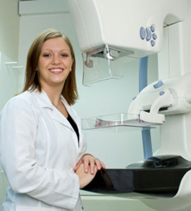
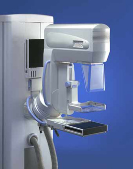
A mammogram is an x-ray examination of the breasts, used to detect and diagnose breast diseases. Mammography is considered the most effective tool for early breast tumor detection. Most medical experts agree that successful treatment of breast cancer often is linked to early diagnosis. Mammography plays a central part in early detection of breast cancers because it can show changes in the breast up to two years before a patient or physician can feel them.
Mammography is a very safe procedure that uses low doses of radiation to produce high quality x-ray images of the breast. Screening mammography is used as a preventive measure for women who have no symptoms of breast disease. A screening mammogram usually involves two views of each breast. Diagnostic mammography involves additional view of the breast, and is used when an abnormality is found during screening, or in women who have breast complaints, such as a breast mass, nipple discharge, breast pain, or skin irritation.
What are the advantages of digital mammography and computer-aided detection?
- With digital mammography, the radiologist reviews electronic images of the breast, using special high-resolution monitors. The physician can adjust the brightness, change contrast, and zoom in for close ups of specific areas of interest. Being able to manipulate images is one of the main benefits of digital technology.
- Another convenience of digital mammography over film-based systems is that it can greatly reduce the need for retakes due to over or under exposure. This potentially saves additional time and reduces your exposure to x-rays.
- Because they are electronic, digital mammography images can be transmitted quickly across a network. Digital images can also be easily stored, copied without any loss of information, and transmitted and received in a more streamlined manner, eliminating dependence on only one set of “original” films.
- Compared with standard film-based mammograms, digital mammograms are more accurate, especially in younger women with dense breast tissue, women under 50 years old, and those who are premenopausal.
Digital mammography has introduced another tool in helping the radiologist detect breast cancer – CAD, or computer-aided detection. CAD systems use computer software to analyze mammograms. They highlight regions of interest, drawing the radiologist’s attention to areas that require additional review. CAD technology basically works like a second pair of eyes, equivalent to having the mammogram double-read by another radiologist. Studies have shown CAD further improves the detection of breast cancer on screening mammograms.
Breast Ultrasound
Ultrasound uses sound waves to generate a picture of the breast tissue. No compression is necessary. Ultrasound is particularly useful in telling cysts from solid masses in the breast. Cysts are very common and totally benign; about half of all women have some cysts in their breasts at some point. Ultrasound is also very helpful in characterizing masses and lumps.
Sometimes we do find a lesion that requires a biopsy to find out what it is. Fortunately, the vast majority of breast biopsies can be accurately performed with a needle and do not require surgery. Depending on the finding, a needle biopsy is performed, using either stereotactic (mammogram), ultrasound, or MRI guidance.
Breast Biopsy
Women who require minimally invasive needle biopsy of the breast can be confident in the services provided by the highly skilled and experienced radiologists at Medical Arts. These imaging controlled procedures may be performed with mammographic (stereotactic), ultrasound, or MRI guidance.
Biopsies are performed under local anesthesia and are outpatient procedures. The incision is small and requires no stitches. There is no deformity of the breast or scarring on mammograms after a needle biopsy, and complications such as serious bleeding or infection are extremely rare.
In most patients, lesions will prove to be benign, therefore making surgery unnecessary. Patients diagnosed with breast cancer by image-guided biopsy will require fewer surgeries than those diagnosed by surgical biopsy. Today, minimally-invasive image-guided breast biopsies have largely replaced surgical biopsies.
Stereotactic-Guided Breast Biopsy. The stereotactic table is specially designed so that you can lie face-down with one breast positioned through a hole in the table. Two digital x-ray images are taken from different angles, allowing the radiologist to precisely localize the area to biopsied. Once the area has been located, the radiologist numbs the area with a local anesthetic, then uses computer guidance for precise needle placement and collection of small tissue samples.
Ultrasound-Guided Breast Biopsy. The radiologist uses ultrasound to locate the area for biopsy and to direct the needle used in collecting breast tissue samples.
Both methods are as accurate as a surgical biopsy and are performed on an outpatient basis, taking less than 40 minutes to perform and requiring no stitches. Furthermore, the patient can resume normal, non-strenuous activities immediately after the procedure is done. The samples are sent to the pathology lab for analysis. Results are usually available within 48 hours.
What is MRI?
Magnetic resonance imaging, or MRI, uses strong magnet and radio waves to provide clear and detailed diagnostic images of internal body organs and tissues. MRI is a valuable tool for the diagnosis of a broad range of conditions, including:
- cancer
- heart and vascular disease
- stroke
- joint and musculoskeletal disorders
MRI allows evaluation of some body structures that may not be as visible with other diagnostic imaging methods.
What are some common uses of MRI?
Imaging of the Musculoskeletal System: MRI is often used to study the knee, ankle, foot, shoulder, elbow, wrist, and hand. MRI is also a highly accurate method for evaluation of soft tissue structures such as tendons and ligaments, which are seen in great detail. Even subtle injuries are easily detected. In addition, MRI is used for the diagnosis of spinal problems including disc herniation, spinal stenosis, and spinal tumors.
MRIImaging of the Heart: MRI of the heart, aorta, coronary arteries, and blood vessels is a tool for diagnosing coronary artery disease and other heart problems. Doctors can examine the size and thickness of the chambers of the heart and determine the extent of damage caused by a heart attack or heart disease.
Imaging for Cancer & Functional Disorders: Organs of the chest and abdomen such as the liver, lungs, kidney, and other abdominal organs can be examined in great detail with MRI. This aids in the diagnosis and evaluation of tumors and functional disorders. In the early diagnosis of breast cancer, MRI is an alternative to traditional x-ray mammography. Furthermore because there is no radiation exposure is involved, MRI is often used for examination of the male and female reproductive systems.
How should I prepare for an MRI?
- Before your MRI exam, remove all accessories including hair pins, jewelry, eyeglasses, hearing aids, wigs, dentures. During the exam, these metal objects may interfere with the magnetic field, affecting the quality of the MRI images taken.
- Notify your technologist if you have:
- any prosthetic joints – hip, knee
- a heart pacemaker (or artificial heart valve), defibrillator or artificial heart value
- an intrauterine device (IUD),
- any metal plates, pins, screws, or surgical staples in your body.
- tattoos and permanent make-up.
- a bullet or shrapnel in your body, or ever worked with metal.
- if you might be pregnant or suspect you may be pregnant.
- if you are claustrophobic. Some patients who undergo MRI in an enclosed unit may feel confined. If you are not easily reassured, a sedative may be administered.
Breast MRI and MRI-guided Breast Biopsy
Breast MRI is an extremely helpful imaging tool in evaluating mammogram abnormalities and identifying early breast cancer, especially in women at high risk. MRI does not replace screening mammography. Instead, it provides a powerful supplementary tool for detecting and staging breasts cancer.
MRI is a technique using a very strong magnet and radio waves to pick up signals from the breast tissue. We use state-of-the-art equipment including a dedicated bilateral breast surface coil. The patient lies face-down within the scanning field for approximately 25 minutes. The primary way that abnormal tissue stands out on MRI is because it gets more blood flow than the remaining tissue. We can detect blood flow by taking images before and after infusion of an intravenous substance (gadolinium) that is easily seen on MRI. We utilize a special computer-assisted detection (CAD) program designed for the processing and interpretation of breast MR images.
Breast MRI is most useful in screening for breast cancer in high risk patients and evaluating the integrity of implants. Breast MRI is also employed in patients with a known breast cancer in whom there is a question about how extensive the disease is.
What is Dexa Scan?
To accurately detect osteoporosis, doctors commonly use DEXA bone densitometry to measure bone mineral density (BMD). DEXA is a quick, painless procedure for measuring bone loss. Measurement of the lower spine and hips are most often done.
Peripheral dual-energy X-ray absorptiometry (P-DEXA) machine, which measures bone density in the wrist or forearm, are portable units that can be used in a doctor's office.
What are some common uses of this procedure?
DEXA bone densitometry is commonly used to diagnose osteoporosis, a condition that often affects women after menopause but may also be found in men. Osteoporosis involves a gradual loss of calcium, causing bones to thin, become more fragile, and more likely to break.
The DEXA test can also assess your risk for developing fractures and is effective in tracking the effects of treatment for osteoporosis and other conditions that can cause bone loss. Bone density testing is recommended for:
- post-menopausal women age 60 or older who have risk factors for developing osteoporosis
- patients with a personal or maternal history of hip fracture or smoking
- post-menopausal women who are tall (over 5 feet 7 inches) or thin (less than 125 pounds)
- men and women who have hyperparathyroidism
- men and women who have been medications that are know to cause bone loss for an extended period of time
How should I prepare for this procedure?
- Refrain from taking calcium supplements for at least 24 hours beforehand.
- Wear comfortable clothing and avoid garments that have zippers, belts or buttons made of metal.
- Let your technologist know if you’ve recently had a barium examination or have been injected with a contrast material for a CT or radioisotope scan.
- Let your technologist know if there is a possibility you are pregnant.
What should I expect during this exam?
Depending on the equipment used and the parts of the body being examined, the test takes between 10 and 30 minutes.
- You may be asked to undress and put on a gown.
- You'll lie on a padded table with an x-ray generator below and a detector (an imaging device) above. It is important that you remain as still as possible during the procedure to ensure a clear and useful image.
- When evaluating bone loss in the spine and hip where most osteoporosis-related fractures happen:
- Spine: During an examination of the spine, your legs will be supported on a padded box to flatten your pelvis and lower (lumbar) spine.
- Hip: The technologist will place your foot in a brace that rotates the hip inward.