
 Nuclear Medicine/PET/CT
What is Nuclear Medicine?
Nuclear medicine, or scan, uses a small amount of a radioactive substance to produce two or three dimensional images of body anatomy and function. The diagnostic images produced by a nuclear scan are used to evaluate a variety of diseases. Sometimes a nuclear scan is combined with a CT scan.
What are some common uses of Nuclear Medicine?
Nuclear medicine images can assist the physician in viewing, monitoring, or diagnosing:
- tumors
- blood flow and function of the heart
- respiratory and blood-flow problems in the lungs
- organ function – of the kidney, bowel, gallbladder and others
How should I prepare for this procedure?
Usually, no special preparation is needed. However, if the exam is done to evaluate the stomach, you may be asked to refrain from eating immediately before the test. If the exam is done to evaluate the kidneys, you may need to drink plenty of water before the test.
What should I expect during this exam?
Although imaging time can vary, the exam generally takes 20 to 45 minutes.
- A radiopharmaceutical, known as a tracer, is usually administered either intravenously or by mouth. What radiopharmaceutical is used and when the imaging will be done - immediately, a few hours later, or even several days after the injection, is dependent upon the type of exam you’re having.
- For most nuclear scans, you will lie down on a table and a nuclear imaging camera will be used to capture the image of the area being examined. The camera is either suspended over or below the exam table or in a large donut-shaped machine similar to a CT scanner. While the images are being obtained, you must remain as still as possible.
- Most of the radioactivity is expelled out of your body in urine or stool. The rest simply disappears through over time.
What will I experience during the procedure?
Although usually done with a small needle, some patients experience some minor discomfort from the intravenous injection, or IV. Also, lying still on the examining table may be uncomfortable for some patients. You will hear low-level clicking or buzzing noises from the machine.
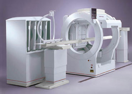
PET/CT
PET/CT combines two machines in one, so that the physiologic activity of PET can be matched with the anatomical information of CT. The two studies are fused, creating one image. The result is greater diagnostic accuracy in localizing abnormalities than PET or CT alone.
PET/CT scanning is performed after injecting radio-labeled sugar (FDG) into an arm vein. After waiting about an hour to allow the sugar to circulate, the scan is started. Since cancerous cells are metabolically more active than normal cells, and therefore use more sugar for energy, they appear as "hot-spots" relative to normal cells. This information, fused with the anatomical CT information, pinpoints the exact location of the abnormality.
PET/CT leads to improved diagnostic confidence and can reduce the need for biopsies and unnecessary surgeries. Diagnoses can be made earlier, with greater accuracy in staging and localization, with more precise treatment and better patient monitoring. Finally, overall patient comfort is increased due to shorter scan times.
PET/CT can be used, as follows, for various types of imaging:
Oncology
- Determine benign from malignant in suspicious areas
- Assess tumor aggressiveness-grading
- Whole body scan for distant metastases
- Early detection of recurrent tumors
- Superior detection of nodal spread
- Monitor success of therapy
Neurology
- Diagnose Alzheimer's disease and other dementias
- Parkinson's disease - diagnosis of movement disorders
Cardiology
PET/CT has a high degree of accuracy for presence or spread of:
- lung cancer
- colorectal cancer
- ovarian cancer
- melanoma
- breast cancer
- brain tumors
- lymphoma
- head and neck tumors
- pancreatic cancer
PET/CT imaging offers fast results with no harm to the patient in the diagnosis, staging and restaging in many areas of oncology. PET/CT has shown to be an asset to many physicians in determining the treatment needed for their patients. Most insurance companies cover PET/CT scan for lymphoma, lung, breast, colorectal and esophageal cancers, head and neck cancer, and melanoma.
PET/CT Patient Preparation
The following high protein/low carbohydrate diet MUST be followed 24 hours prior to your PET/CT scan:
Foods to AVOID :
|
|
Foods that MAY be consumed:
- Plain meat
- poultry
- fish (no breading),
- cheese
- eggs
- bacon
- sausage
- only green vegetables
Drink water with every meal the day before the test. Three to four glasses of water should be consumed the day of the test.
DO NOT DRINK LIQUIDS THROUGH A STRAW
NO EXERCISE 48 HRS PRIOR TO TEST
You are required to fast 6-8 hrs prior to your appointment time. You may take medications with water. Dress comfortably – loose clothing, no metal.
You can expect to be at Medical Arts for approximately two hours.
What should I expect during this exam?
You receive an intravenous (IV) injection of the radioactive substance.
The radioactive substance will then take approximately 30 to 90 minutes to travel through your body and be absorbed by the tissue under study. During this time, you will be asked to rest quietly and avoid significant movement or talking, which may alter the localization of the administered substance.
You will be positioned on the PET scanner table and be asked to lie still during your exam. Scanning takes 30 to 45 minutes.
Some patients who are being evaluated for heart disease may undergo a stress test in which PET scans are obtained while they are at rest, and again after undergoing the administration of a pharmaceutical to alter the blood flow to the heart.
Usually, there are no restrictions on daily routine after the test. You should drink plenty of fluids to flush the radioactive substance from your body.